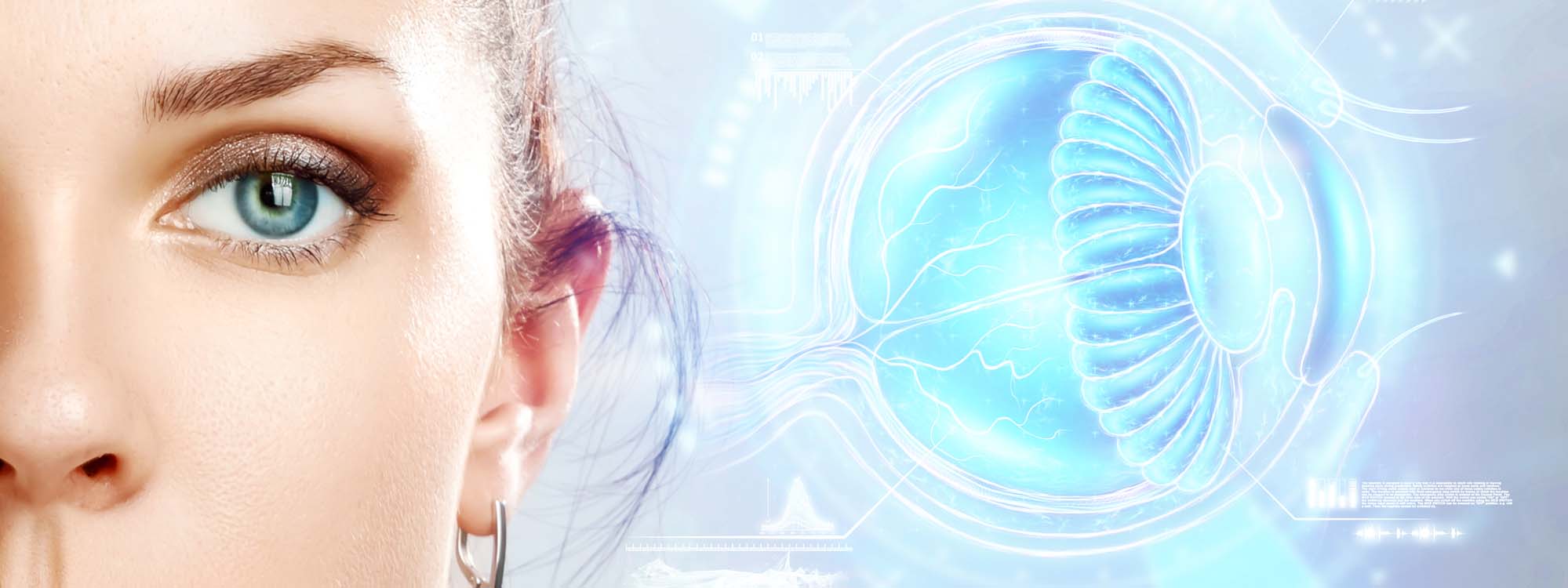
Laser Surgery (Photocoagulation) Treatment
Photocoagulation is a type of laser surgery used to treat detachment of the retina. This treatment involves the use of an argon laser that converts a high-intensity beam of light into heat that seals the tear in the retinal tissue. Laser therapy can also prevent the growth of abnormal blood vessels, a common side effect of retinal detachment. Photocoagulation for the treatment of a detached retina can prevent further vision loss and retinal abnormalities.
Retinal Types
There are three main types of retinal detachment. The first, rhegmatogenous detachment develops due to age because the fluid-filled vitreous body in the middle of the eye shrinks as we age. This can cause the retina to detach from the vitreous body and cause visual disturbances.
The second type of traction is called retinal detachment. This common occurs in people with diabetes because of glucose-mediated inflammation combined with poor circulation. The third type of retinal detachment is called exudative attachment and is the result of a buildup of fluid between the retina and the choroid, a structure located under the retina. When fluid builds up, it can cause the retina to detach. This type of separation is often caused by cancer or inflammatory disorders.
Photocoagulation can be used as a treatment for all three types of retinal detachment. This type of treatment uses an argon laser. This laser is narrowly focused into a beam of light, which is then directed to a portion of the detached retina at the back of the eye. The light beam focuses on the specific spot where the retina detaches. When the light beam reaches the retina, the light is absorbed by the cells and then converted into heat energy. Healing seals the detached retina. This treatment usually takes thirty minutes or less.
Why is Photocoagulation Applied?
Laser photocoagulation is applied in the treatment of various choroidal and retinal tumors. Choroidal nevi are the most common intraocular tumor. Its incidence in the community has been reported to be 6% on average. Vision loss occurs in approximately 10% of choroidal nevi. Causes of vision loss can be listed as subretinal hemorrhage due to choroidal neovascularization, subretinal fluid and pigment epithelial detachment. Laser photocoagulation is used to dam the subretinal fluid originating from the choroidal nevus or to close the leaking foci within the tumor. In choroidal hemangioma, which is also a common eye tumor in adulthood, symptomatic (in terms of vision) improvement is achieved as a result of the disappearance of subretinal fluid by forming adhesions between the retina and the retinal pigment epithelium (coagulation necrosis effect). Laser photocoagulation is the most commonly used method in retinal capillary hemangiomas, especially in lesions smaller than 3 mm and not accompanied by subretinal fluid. With laser photocoagulation, tumor regression, dilatation and tortuosity reduction in feeding and draining vessels, and regression in hard exudation are observed. Laser photocoagulation is repeated at intervals of 4-6 weeks until the expected results from the treatment are seen.
How should the patient be prepared for treatment?
To prepare for photocoagulation therapy, the patient is given eye drops to numb the eye and dilate the pupil. Treatment is usually painless, but eye drops are necessary as some patients are sensitive to laser light. When the treatment is over, the patient can leave immediately. Due to the increased sensitivity to light, the eye should be kept closed for several hours. In addition, the patient should adjust the treatment home, because the drugs given before the treatment can reduce the ability to drive.
